Objective: We aimed to investigate the regional distribution of grey matter (GM) and white matter (WM) atrophy in patients with relapsing-remitting (RR) MS and their relationship with gender, clinical findings, and T2 lesions.
Methods: Clinical and magnetic resonance imaging assessments were obtained from 78 patients with RRMS and 88 controls. GM and WM atrophy were estimated using voxel-based morphometry (SPM8).
Results: Patients with RRMS experienced atrophy of the deep GM nuclei, and several cortical regions mainly located in the fronto-parietal lobes. WM atrophy involved posterior (at the back) regions of the brain, including the cerebellum and brainstem. Compared with men, affected women showed atrophy of several WM tracts, while gender did not impact GM atrophy. Disease duration > 5 years was associated with atrophy of the thalami and inferior frontal gyrus, while WM atrophy was already prominent in patients with disease duration ≤ 5 years. Expanded Disability Status Scale score > 3.0 was associated with diffuse WM atrophy (shrinkage) and basal ganglia and precentral gyrus atrophy. T2 lesions were marginally associated to focal atrophy.
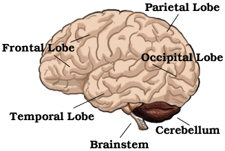

This is a descriptive study, follow the links to see what each bit of the brain does. This study reports on brain shrinkage during MS. The problem of using MRI to assess shrinkage is that this can be masked by fluid accumulation.